Transcranial magnetic stimulation to frontal cortex, unlike occipital stimulation, does not disrupt exogenous attention
Orienting covert attention to a target location improves performance across a wide array of visual tasks [Carrasco, 2011; Carrasco & Barbot, 2014]. Whereas fMRI studies have identified partially overlapping frontoparietal networks underlying endogenous (voluntary) and exogenous (involuntary) covert attention, these correlational methods cannot establish whether a given region is functionally necessary. Prior neurostimulation studies have established that the early visual cortex (V1/V2) is critical for exogenous [Fernández & Carrasco, 2020; Lee et al., 2024] but not endogenous attention [Fernández et al., 2023], whereas the right frontal eye field (rFEF+) is critical for endogenous attention [Fernández et al., 2023]. Here, we used a combined psychophysical-TMS protocol to investigate whether rFEF+ is also required for exogenous attention. Participants performed an orientation discrimination task, in which a peripheral cue (valid, neutral, or invalid) preceded the target and distractor stimuli. We applied two successive TMS pulses to rFEF+ during stimulus presentation and measured contrast-response functions (CRFs) to quantify perceptual sensitivity (d') across all attention cueing and stimulation conditions. When the distractor was stimulated, exogenous attention yielded a characteristic response gain—with performance benefits for valid cues and costs for invalid cues at high contrast levels. Crucially, this response gain was entirely preserved when the target was stimulated. This pattern contrasts with previous findings demonstrating that TMS to V1/V2 eliminates exogenous attentional effects at the stimulated location. These results indicate that rFEF+ is not necessary for exogenous attention. Together with previous studies [Fernández & Carrasco, 2020; Lee et al., 2024; Fernández et al., 2023], these results complete a double dissociation: rFEF+ is critical for endogenous but not exogenous attention, whereas V1/V2 is critical for exogenous but not endogenous attention. These findings reveal a distinct causal cortical architecture for voluntary and involuntary spatial attention, suggesting that they rely on different cortical scaffolds.
Interactive Demo Disclaimer
Please be advised that this demo is ONLY a conceptual illustration designed to demonstrate the experimental task sequence. It is NOT a calibrated reproduction of the laboratory experiment.
Note: Parameters in this demo—including spatial aspects, luminance/colors, stimulus parameters, timing—are optimized for standard web displays and differ from the rigorous, hardware-calibrated settings used in the actual study.
For precise experimental specifications, please consult the Methods section of the full paper.
Do you agree to proceed for illustrative purposes only?
Results
Naka–Rushton functions

Mean parameter estimates
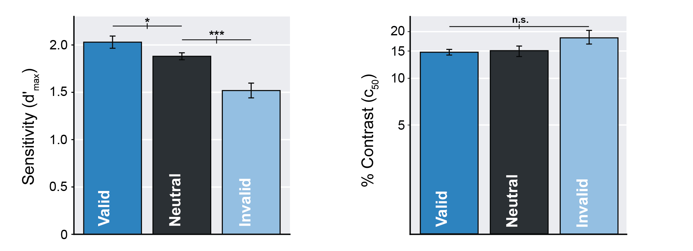
Error bars represent ±1 SEM. ***P≤0.001, *P≤0.05
Microsaccades Analysis

Group-averaged microsaccade rate relative to cue onset, split for target location (target stimulated, distractor stimulated). Vertical gray lines denote stimulus timing: cue onset, stimulus onset, and response cue onset, and vertical dashed black lines denote TMS pulses.
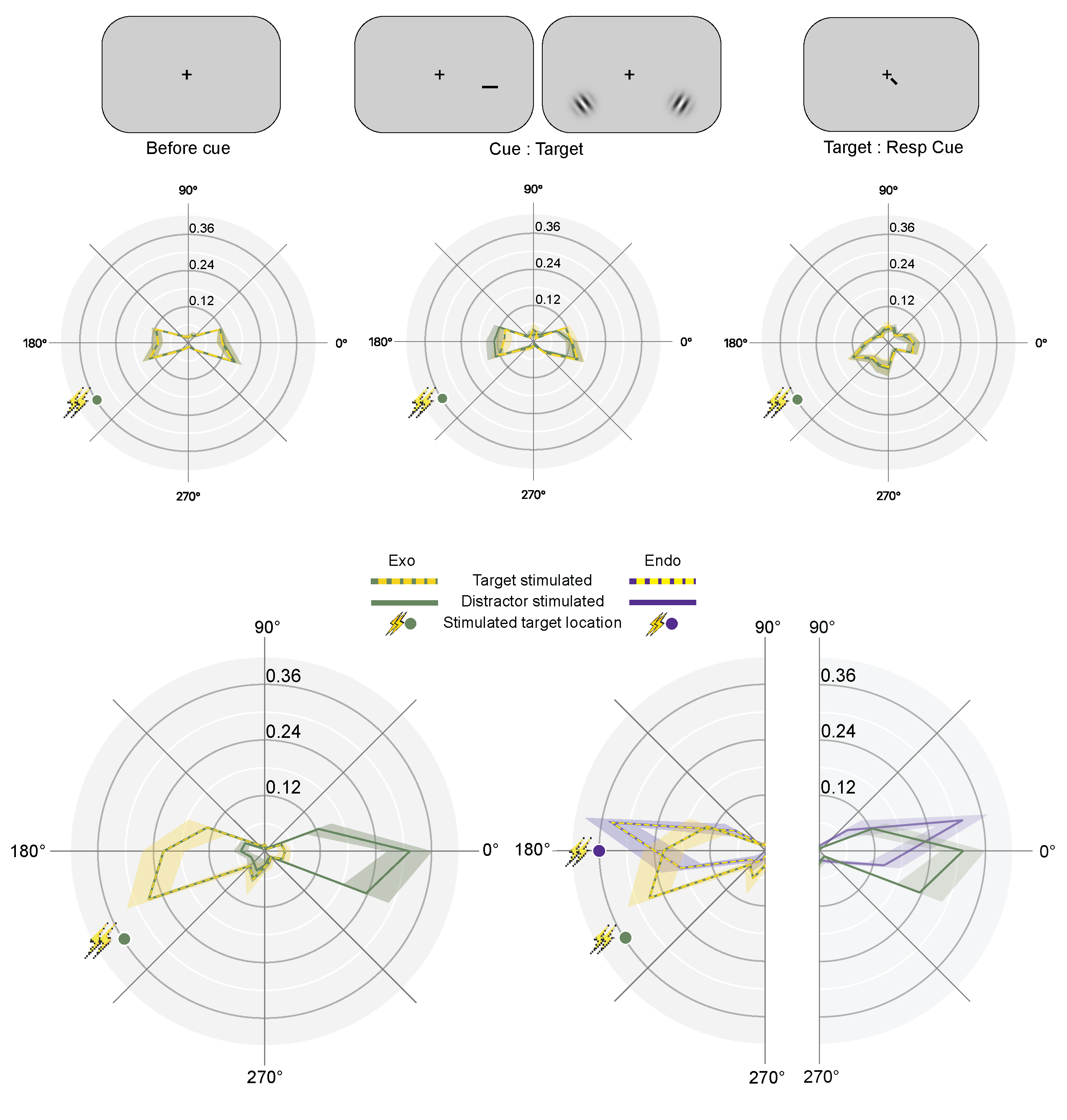
Upper panel: Normalized group-averaged polar angle microsaccade frequency before the cue (left panel), from cue onset to target onset (middle panel), and between target onset and response cue (right panel). Lower panel: Normalized group-averaged polar angle microsaccade frequency after response cue onset (up to response) following rFEF+ TMS, split for target location (target-stimulated and distractor-stimulated conditions); current rFEF+ Exo data (left panel) and comparison with rFEF+ Endo data reported in Fernández, Hanning & Carrasco (2023) (right panel). Dots indicate target location in stimulated hemifield (green: Exo rFEF+; purple: Endo rFEF+). Colored, shaded areas in all panels indicate ±1 SEM.
Simulated cortical excitability
Estimated E-field for the rFEF+ using the simNIBS toolbox.